|
|
Calcium Hydroxyapatite Deposition Disease
HADD
General Considerations
- Calcium phosphate crystals are deposited in the form of calcific tendinitis, other periarticular hydroxyapatite deposition, and hydroxyapatite-induced arthritis
- May be primary, such as calcific tendinitis and bursitis, or secondary, as in dystrophic calcification in chronic renal disease or collagen vascular diseases

- Most common in middle aged men, slightly greater than women
- The shoulder is the most common site
Clinical Findings
- Recurrent pain, either chronic or acute
- Limitation of motion
- Asymptomatic deposits in the shoulder are common
- About half of patients with shoulder deposits have them in both shoulders
- Most are in the supraspinatus tendon near the greater tubercle
- Hip (external rotators), elbow wrist and knee follow in that order
Imaging Findings
- Homogeneous, sharply defined, well-circumscribed amorphous collections of calcium density without trabeculation
- Most deposits are ovoid in shape
- Most are near the joint but some can be found some distance from a joint
- Multiple deposits are common both in joints and surrounding tendons
- Over time, the deposit may become liquefied
- The deposit may then rupture into the bursa or be subursal
- Finally, disappearance of calcification is common
- MR Findings
- Low signal focus on all sequences
- Adjacent soft tissues may be hyperintense
- May have “blooming” of deposit
- Calcification can also be intra-articular, as in shoulder joint
- Usually occurs in elderly women (Milwaukee shoulder)
Differential Diagnosis
Treatment
- Conservative
- Steroid injection
Prognosis
- Usually self-limiting with resolution of both clinical and imaging findings

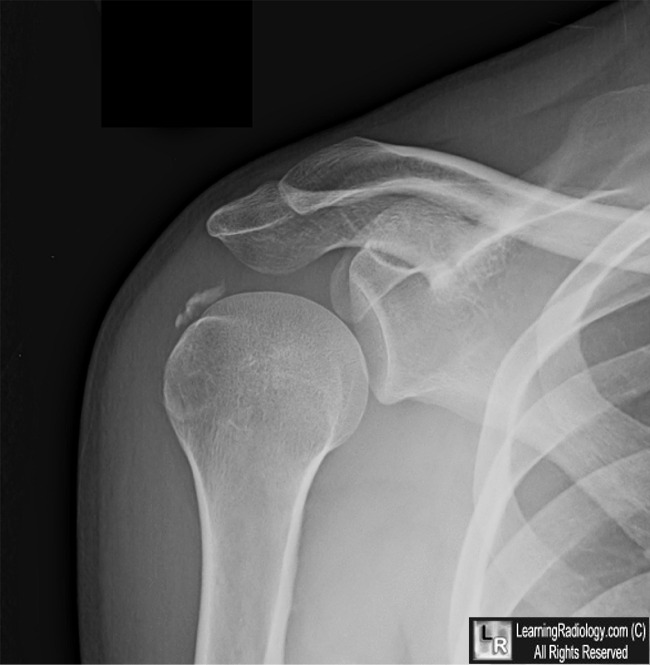
Hydroxyapatite Deposition Disease. There are well circumscribed, amorphous calcifications not containing trabeculae adjacent to and paralleling the greater tubercle of the humerus (white arrow).
For this same photo with the arrows, click here
For more information, click on the link if you see this icon 
Calcium Hydroxyapatite Deposition Disease. C Hayes and W Conway. November 1990 RadioGraphics, 10, 1031-1048.
|
|
|
